Advancements in technology and diagnostics tools have always been vital to enhancing patient care. Among these, digital X-rays have become a vital solution in the field of dentistry, transforming the way dental professionals diagnose and treat various conditions. As part of our commitment to advanced dental care, we at Julie Ann Valde, DDS, prioritize the implementation of the latest technology, including digital X-rays. In this comprehensive guide, we’ll take a deep dive into the world of digital X-rays, exploring their workings, numerous benefits, and tips for optimizing your digital X-ray experience; and we'll address common questions and safety concerns that some patients may have.
Digital X-Rays: Revolutionizing Dental Diagnostics
Traditional film-based X-rays have been eclipsed by digital X-rays in the realm of dental diagnostics. The transformation is in the use of digital sensors instead of film to capture highly detailed images of the teeth, gums, and jaw structures. These images can be instantaneously viewed on a computer screen, thus optimizing the diagnostic process in terms of efficiency, accuracy, and safety.
The Benefits of Digital X-Rays
The shift from film-based to digital X-rays has revolutionized dental diagnostics by offering many key advantages.
Enhanced Images for Superior Diagnosis
Digital X-rays have the capability to produce high-resolution images that capture more detail than traditional film X-rays. This enhanced image quality paves the way for a comprehensive analysis, enabling dentists to formulate accurate diagnoses and devise effective treatment plans.
Enlargement Options: A Closer Look
In the digital format, images can be easily enlarged, allowing dentists to focus closely on specific areas. This feature is particularly useful in the early identification of potential issues which may go unnoticed with traditional X-rays.
Immediate Viewing: Speeding Up Diagnostics
Gone are the days when patients had to wait for films to be developed. Digital X-rays are instantly available for viewing, expediting the diagnosis process. This immediacy allows dentists to swiftly discuss issues with patients and start necessary treatments without unnecessary delay.
Improved Diagnostics: Spotting the Unseen
The clarity provided by digital X-rays facilitates the detection of hidden dental issues. These can range from decay and bone infections to gum disease, tumors, and other abnormalities that might not be visible during a conventional visual examination or with a traditional X-ray.
Time and Cost Savings: An Economical Approach
With digital X-rays, the early detection of dental problems minimizes the need for more invasive and expensive treatments. In the long run, this results in significant time and cost savings for patients.
Reduced Radiation: Safety First
One of the primary benefits of digital X-rays is their ability to reduce radiation exposure by up to 70%, as compared to traditional X-rays. This aspect makes digital X-rays a much safer diagnostic method.
Electronic Storage & Easy Access: The Digital Advantage
Digital X-rays are stored electronically, providing easy access and efficient sharing options. These digitized records can be shared with other healthcare providers, insurance companies, or even the patient themselves. This convenience streamlines communication and expedites insurance claims.
Environmentally Friendly: A Sustainable Choice
Digital X-rays also contribute to environmental conservation. Unlike traditional methods, they don’t require harmful chemicals for developing films, thus reducing their ecological impact. At Julie Ann Valde, DDS we are committed to protecting our environment, so we are proud to use this technology to reduce our footprint.
The Technology behind Digital X-Rays
Digital X-ray technology, also known as digital radiography, has revolutionized the field of dental imaging. But what exactly makes digital X-rays tick?
The Digital Sensor: The Heart of the Technology
At the core of digital X-ray technology is a digital sensor. This small device is placed in the patient's mouth to capture the images. Unlike traditional film that must be developed, these digital sensors directly capture and convert X-ray energy into electronic signals.
The Software: Making Sense of the Data
The signals from the digital sensor are sent to a computer where they are interpreted and converted into a visual image by dedicated software. The computerized nature of this process allows for a variety of image enhancement tools to be used, helping dental professionals to spot any issues that might require attention.
The Monitor: Immediate Viewing
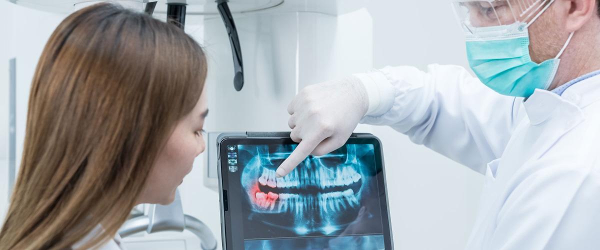
The final images are instantly viewable on a computer monitor. This immediacy not only saves valuable time but also aids in patient communication, as our dentist can discuss the images and any identified issues with the patient right away.
Ensuring Safety with Digital X-Rays
Despite the significant reduction in radiation exposure, digital X-rays do still involve some radiation. At Julie Ann Valde, DDS, patient safety is paramount. We adhere to strict safety protocols and perform X-rays only when necessary. Patients with special conditions, including pregnant women, should discuss the potential risks with their dentist before undergoing any form of radiography.
Types of Digital Dental X-Rays and Their Uses
Digital dental X-rays come in different types, each designed to capture specific views of your teeth and mouth. The choice of which type to use depends on what your dentist is trying to discover or monitor.
Bitewing X-Rays
Bitewing X-rays are named after the small plastic bite tabs that patients hold onto while the X-ray images are captured. These X-rays are used to examine the upper and lower back teeth in a single view. They help the dentist identify cavities between teeth and changes in bone density caused by gum disease. They also aid in the assessment of the proper fit of a crown or other restorations.
Periapical X-Rays
Periapical X-rays focus on one or two complete teeth, from the root to the crown. These X-rays are typically used when a dentist wants a detailed view of a particular tooth or teeth. They can help identify any abnormalities in the root structure and surrounding bone structure, diagnose an abscess, or monitor the progress of a particular dental treatment.
Panoramic X-Rays
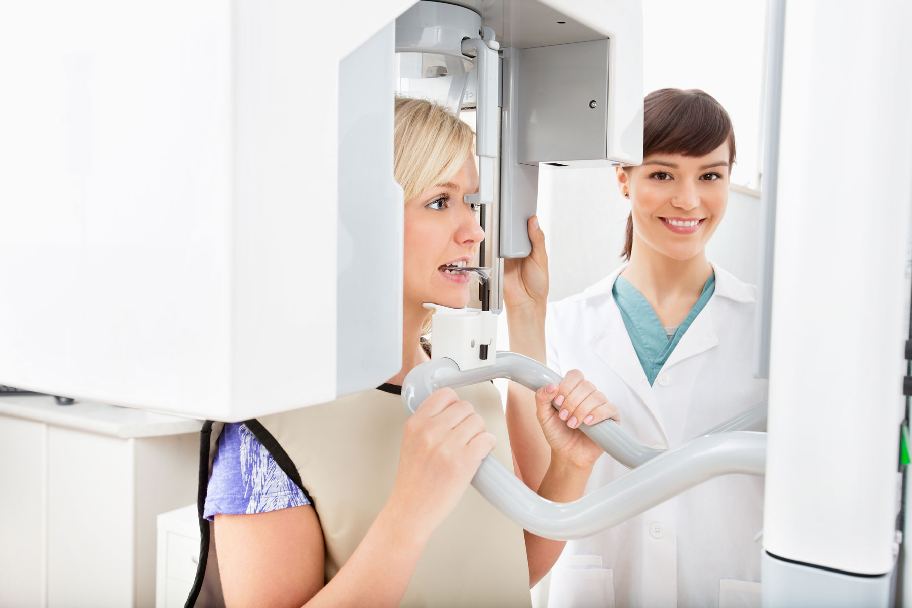
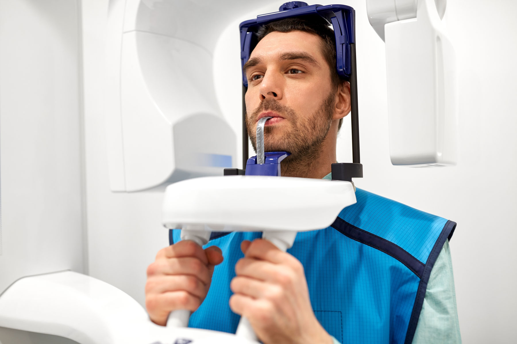
Panoramic X-rays provide a broad view of the entire oral cavity. This type of X-ray captures images of the teeth, upper and lower jawbones, sinuses, and temporomandibular joints (TMJ) in one single image. Because of the comprehensive view it provides, it is typically used for planning treatments such as implants, braces, dentures, or extractions. It is also used to identify issues such as fractures, cysts, tumors, bone irregularities, and infections in the jaw.
Optimizing Your Digital X-Ray Experience
To maximize the benefits from your digital X-ray experience, open and proactive communication with your dentist is key. Ask questions, understand the process, and ensure you’re aware of the potential outcomes. Following all given instructions and promptly reporting any changes in your dental health will further enhance your digital X-ray experience.
Why Choose Julie Ann Valde, DDS for Your Dental Care?
At Julie Ann Valde, DDS, we are committed to providing the ultimate in dental care. Our highly trained dentists use digital X-rays to diagnose and treat a diverse array of dental conditions. Beyond digital X-rays, our comprehensive services span from oral cancer detection to dental hygiene advice and cosmetic services. Julie Ann Valde, DDS is your reliable partner for dental care.
We use digital X-rays as part of our routine dental wellness exams. Schedule your wellness exam today and let us take care of you!


